· News · 15 min read
Out now in Immunity: PD-L1 on FDCs shapes germinal centers
Almost seven years after finding PD-L1 on human follicular dendritic cells, we can now explain how stromal PD-L1 shapes germinal centers.
In early 2019, I contacted my PhD advisor, Mike Carroll, with an observation: human follicular dendritic cells (FDCs) express PD-L1 and PD-L2. We reported that finding in the Journal of Experimental Medicine in 2021. Almost seven years (and a few CXCL13-Cre, PD-L1-flox mice) later, we can finally answer the next question: what does PD-L1 do on FDCs?
Our new paper in Immunity, led by Elliot Akama-Garren, shows that PD-L1 on germinal-center stromal cells promotes humoral immunity. It does this, in part, by limiting a pathway in which a subset of T follicular helper cells (Tfh) promotes the development of regulatory follicular T cells (Tfr). The work combines a large cross-species stromal-cell analysis with conditional mouse genetics, single-cell and bulk RNA sequencing, imaging, autoimmune models, and analyses of human tumor datasets.
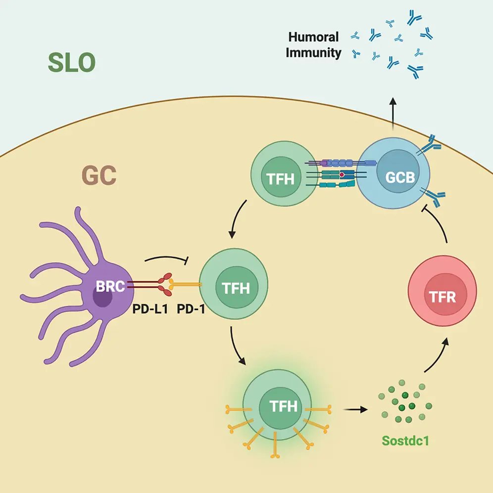
Graphical Abstract from the paper: PD-L1 on FDCs restrains a subset of Tfh cells, limiting Tfr differentiation and supporting germinal-center B-cell responses.
The question: what does PD-L1 do in a germinal center?
PD-L1 is usually introduced as an immune brake. By binding PD-1, it can dampen T-cell activation. That role is central to cancer immunotherapy, where antibodies that block PD-1 or PD-L1 can restore some antitumor immune responses.
But PD-L1 is expressed by different cell types, and its effects depend on the cells it interacts with. In a germinal center (a temporary structure in lymph nodes, spleen, and other lymphoid tissues) B cells compete for antigen and for help from T follicular helper (TFH) cells. T follicular regulatory (TFR) cells help regulate this response. The balance among these cells influences the size, duration, and output of the germinal center.
Previous studies that removed PD-1 or PD-L1 throughout the body had produced seemingly conflicting results for follicular T cells and antibody responses. Such whole-animal experiments affect many cell types at once, making it difficult to tell whether a change originates in B cells, dendritic cells, follicular T cells, or the stromal cells that organize the tissue. We therefore focused on a specific, previously underexplored source of PD-L1: the stromal cells of the B-cell zone, especially FDCs, which are part of the B cell zone reticular cells.
Mapping PD-L1 across lymphoid stromal cells
The first step was to establish which stromal cells express PD-L1. The team generated new single-cell RNA-sequencing data from human tonsils and mouse Peyer’s patches and integrated these with published datasets from multiple lymphoid tissues. Altogether, the analysis included 186 samples and more than 519,000 cells. It resolved several stromal populations, including FDCs, T-cell-zone reticular cells (TRC), and marginal reticular cells (MRC), and identified substantial diversity among FDCs themselves. Despite this diversity, PD-L1 expression was shared across human and mouse FDC populations. FDCs also expressed PD-L2, though less frequently.
The RNA data were followed up at the protein level. Microscopy and flow cytometry confirmed PD-L1 on mouse FDCs in tissue. After immunization, FDC PD-L1 rose rapidly and then returned toward baseline roughly 10-15 days later. That timing was interesting: it suggested that FDC PD-L1 is dynamically regulated during the same period in which germinal centers develop and then contract.
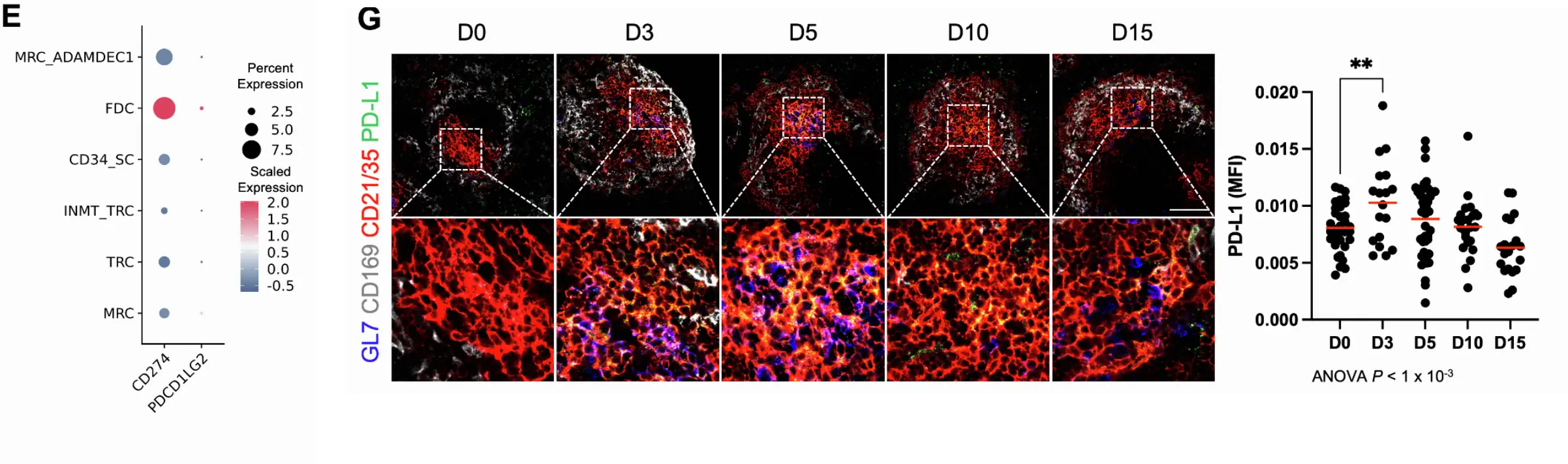
(E) Dot plot comparing the percent of cells and average expression of CD274 and PDCD1LG2 in individual stromal cell clusters from scRNA-seq. (G) Confocal microscopy (left) and quantification (right) of immunofluorescence of GL7, CD169, CD21/35, and PD-L1 expression in mouse spleens on indicated days after immunization with SRBC. Scale, 100 µm. P-value computed using two-tailed Student’s t-test. *, p<0.05; **, p<0.01. Red horizontal lines represent mean. Data are representative of at least two independent experiments.
What happened when stromal PD-L1 was removed?
For the first genetic test, the team crossed Cxcl13-Cre mice with Pdl1 floxed mice. This removed PD-L1 from CXCL13-expressing B-cell-zone reticular cells. Importantly, CXCL13 is not exclusive to FDCs: this model affects FDCs as well as other CXCL13-expressing reticular cells, including some T-cell-zone and marginal-zone stromal cells. So this experiment tests B-cell-zone reticular-cell PD-L1 rather than FDCs alone.
After immunization with the model antigen NP-OVA, mice lacking PD-L1 in these cells had fewer germinal-center B cells and antibody-secreting cells. They also had fewer antigen-specific anti-NP IgG antibodies. Meanwhile, TFR cells were more frequent and showed increased proliferation. The overall frequency of follicular T cells was not reduced, indicating that the result was not simply a loss of the follicular T-cell compartment; instead, the balance shifted toward TFR cells. The mice retained follicular architecture and had similar baseline T- and B-cell numbers, making a general defect in lymphocyte development an unlikely explanation.
That is the first key result: stromal PD-L1 supports the germinal-center response and antibody production while limiting the expansion of TFR cells.
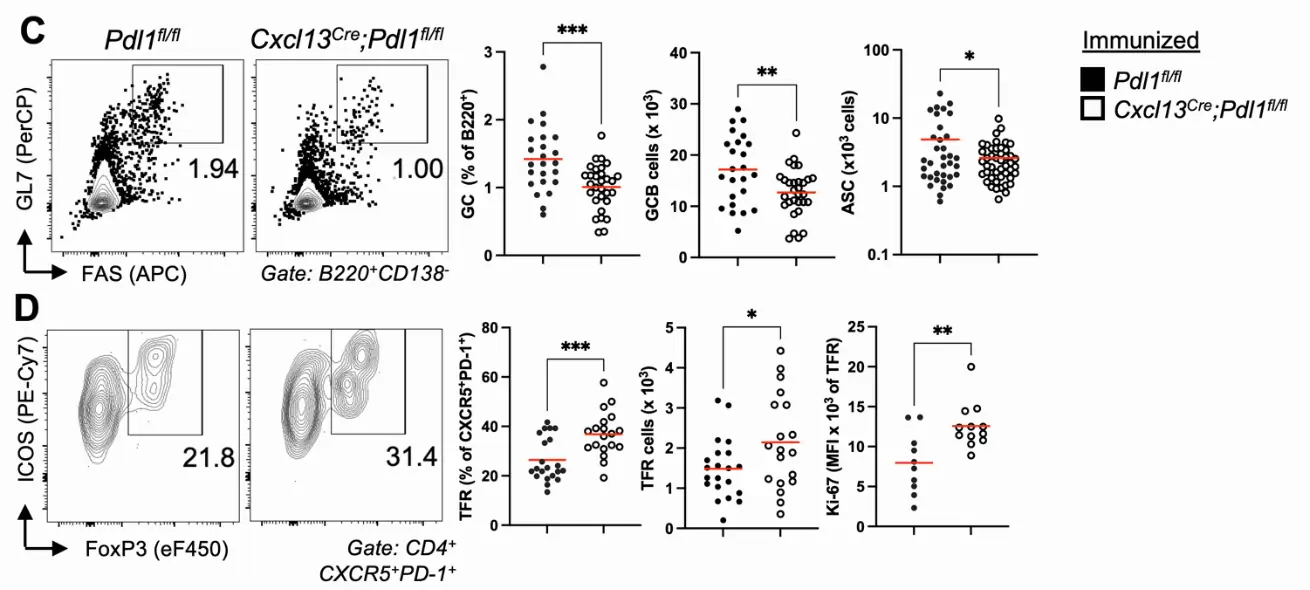
(C) Flow cytometry (left) and quantification (right) of GC (germinal center, B220+CD138-GL7+FAS+) B cell frequency and total ASCs (antibody secreting cells, CD138+) in draining lymph nodes from Pdl1fl/fl (n ≥ 10) or Cxcl13Cre;Pdl1fl/fl (n ≥ 12) mice 10 days after secondary immunization with NP-OVA. (D) Flow cytometry (left) and quantification (right) of TFR (T follicular regulatory, CD4+CXCR5+PD-1+FoxP3+ICOS+) cell frequency and Ki-67 expression.
Testing the role of FDC PD-L1 in autoimmunity
The team (Elliot) next asked whether the same pathway matters in autoreactive germinal centers. They used 564Igi mice, which carry a knock-in autoreactive B-cell receptor and spontaneously develop autoantibody responses. FDCs in these mice expressed more PD-L1 than FDCs in control mice.
When PD-L1 was deleted from CXCL13-expressing reticular cells in the 564Igi model, the mice had fewer germinal-center B cells, antibody-secreting cells, and memory B cells, alongside more numerous and more proliferative TFR cells. Several measures of autoantibody activity also fell, including serum anti-single-stranded DNA antibodies, antinuclear antibody staining, and antibody deposition in kidney glomeruli.
A second experiment asked whether PD-L1 affects the loss of B-cell tolerance that can occur inside autoreactive germinal centers. The researchers made mixed bone-marrow chimeras containing autoreactive 564Igi B cells together with congenically marked, initially non-autoreactive B cells. In this model, non-autoreactive B cells can enter autoreactive germinal centers and expand, a process associated with loss of tolerance and epitope spreading. Removing stromal PD-L1 reduced the dominance of these incoming B cells and reduced autoantibody measures.
Because the Cxcl13-Cre model targets more than FDCs, the team used a second genetic strategy to strengthen the FDC-specific conclusion. In Cr2-Cre chimeras, PD-L1 deletion was restricted to the radiation-resistant FDC compartment, while the hematopoietic cells and other stromal cells retained PD-L1. These mice showed a similar pattern: fewer germinal-center B cells and antibody-secreting cells, more TFR cells and TFR proliferation, fewer memory B cells, and reduced entry of non-autoreactive B cells into autoreactive germinal centers. Together, the two genetic approaches support a specific role for FDC PD-L1 in promoting germinal-center activity and, in these mouse models, contributing to the breakdown of B-cell tolerance.
[Figure idea: depict the mixed-chimera design and the reduced invasion of autoreactive germinal centers by congenically marked non-autoreactive B cells when FDC PD-L1 is absent.]
A subset of TFH cells may connect PD-L1 to TFR cells
The next question was mechanistic: how can PD-L1 on stromal cells change the balance between TFH and TFR cells?
Single-cell RNA sequencing of more than 19,000 TFH cells identified several transcriptional states. One subset expressed high levels of Sostdc1 and Pdcd1, the gene encoding PD-1. As TFH clones became larger, Sostdc1 expression tended to be higher. Trajectory and RNA-velocity analyses were consistent with Sostdc1-producing cells representing a later TFH state. The researchers also sorted TFH cells from autoimmune mice based on PD-1 levels and confirmed that cells with higher PD-1 expressed more Sostdc1. Following immunization, TFH-cell Sostdc1 rose over time, alongside the development of TFR cells and the later decline of germinal-center B cells and antibody-secreting cells. [12]
This fits with previous work showing that Sostdc1 can promote TFR-cell differentiation. In the current study, removing stromal PD-L1 increased Sostdc1 expression in TFH cells. Bulk RNA sequencing showed that these TFH cells adopted a broader Sostdc1-associated gene-expression program, and RNAscope imaging confirmed more Sostdc1-expressing TFH cells in germinal centers. The number of BCL6-positive follicular T cells did not simply increase, pointing to a shift in TFH-cell state rather than a general expansion of all follicular T cells. [12] [13]
The team then tested whether Sostdc1 was functionally involved. Mice deficient in Sostdc1 had stronger germinal-center responses after immunization, fewer TFR cells, and lower TFR proliferation. More directly, removing Sostdc1 from TFH cells in the PD-L1-deficient autoimmune model restored spontaneous germinal-center formation and reduced TFR cells. This rescue experiment makes the Sostdc1-producing TFH population more than a correlate: it supports a causal role for this pathway in the germinal-center phenotype caused by loss of stromal PD-L1. [13]
The model emerging from these experiments is that PD-L1 on FDCs engages PD-1 on a subset of TFH cells and restrains their Sostdc1 program. With less Sostdc1, fewer TFR cells develop or proliferate, leaving more opportunity for productive TFH–B-cell collaboration. The paper also finds that PD-1 can act cell-autonomously to limit follicular T-cell activation, so its biology is not simply “inhibitory” or “stimulatory.” The effect depends on which cell expresses PD-1 or PD-L1 and on the particular interaction being measured. [12] [13]
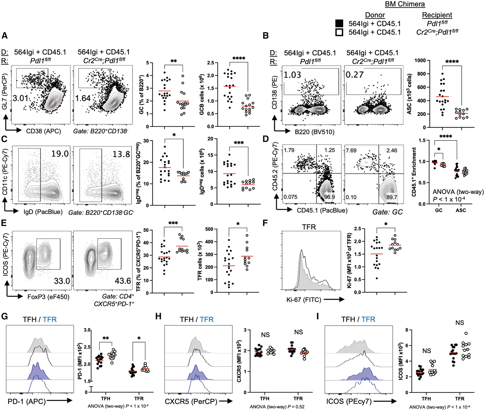
(A–F) Mixed bone marrow chimeras were made by reconstituting irradiated Pdl1fl/fl (n ≥17) or Cr2Cre;Pdl1fl/fl (n ≥12) mice with 1p 564Igi (CD45.2) and 2p CD45.1 bone marrow and analyzed 10 weeks after reconstitution. (A) Flow cytometry (left) and quantification (right) of GC (B220+CD138–GL7+CD38–) B cell frequency in spleens. (B) Flow cytometry (left) and quantification (right) of total ASCs (antibody-secreting cells, CD138+) in spleens. (C) Flow cytometry (left) and quantification (right) of IgD– memory B cell frequency from flow cytometry of spleens. (D) Flow cytometry (left) and quantification (right) of CD45.1 and CD45.2 GC B cell and ASC frequency in spleens. Enrichment was calculated by dividing the CD45.1 frequency in the indicated population by the CD45.1 frequency among follicular B cells (B220+CD138–IgD+GCneg). (E) Flow cytometry (left) and quantification (right) of TFR (CD4+CXCR5+PD-1+FoxP3+ICOS+) cell frequency in spleens. (F) Flow cytometry (left) and quantification (right) of TFR expression of Ki-67 in spleens. (G–I) Flow cytometry (left) and quantification (right) of (G) PD-1, (H) CXCR5, or (I) ICOS expression in TFH and TFR cells in spleens. (J–K) Mixed bone marrow chimeras were made by reconstituting irradiated Pdl1fl/fl (n ≥16) or Cxcl13Cre;Pdl1fl/fl (n ≥12) mice with 1p 564Igi (CD45.2) and 2p CD45.1 bone marrow and analyzed 10 weeks after reconstitution. (J) Confocal microscopy of immunofluorescence of CD4, GL7, CD169, and PD-1 or B220, GL7, CD45.2, and CD45.1 expression in spleens; or DAPI, IgG, and Phalloidin expression from HEp-2 cells stained with sera from Pdl1fl/fl (n = 20) or Cxcl13Cre;Pdl1fl/fl (n = 8) autoimmune chimeras. Scale, 200 μm (spleen) or 50 μm (HEp-2). (K) Quantification of average number of GCs per follicle per mouse (left) or average GC size per mouse (right) in spleens from Pdl1fl/fl or Cxcl13Cre;Pdl1fl/fl autoimmune chimeras. Each point represents the average of multiple fields of view with follicles defined by CD169 expression and GCs by GL7 expression per mouse. p value computed using two-tailed Student’s t test for parametric two-way comparisons or two-tailed Mann-Whitney test for non-parametric comparisons. *p < 0.05; **p < 0.01; ***p < 0.001; ****p < 0.0001. Data are representative of at least three independent experiments.
Watching germinal-center interactions in real time
The study also tested whether the altered T-cell balance was reflected in the behavior of cells inside living lymph nodes. Using intravital multiphoton microscopy, the team tracked fluorescently labeled B and T cells in germinal centers.
Without PD-L1 on B-cell-zone reticular cells, germinal-center B cells moved more slowly and with more meandering, while TFH cells moved faster and with more meandering. TFR cells moved more slowly. The researchers then used the distances between tracked cells to identify interactions. Stable contacts between germinal-center B cells and follicular T cells formed less often and lasted for less time when stromal PD-L1 was absent.
These movies provide a cellular-level link to the flow-cytometry results: when TFR cells are more abundant and proliferative, B-cell–TFH contacts become less stable. The imaging does not, on its own, prove that altered contact duration is the only reason for the weaker antibody response. But it gives a compelling view of how changing the follicular T-cell balance can alter the physical interactions on which germinal-center selection depends.
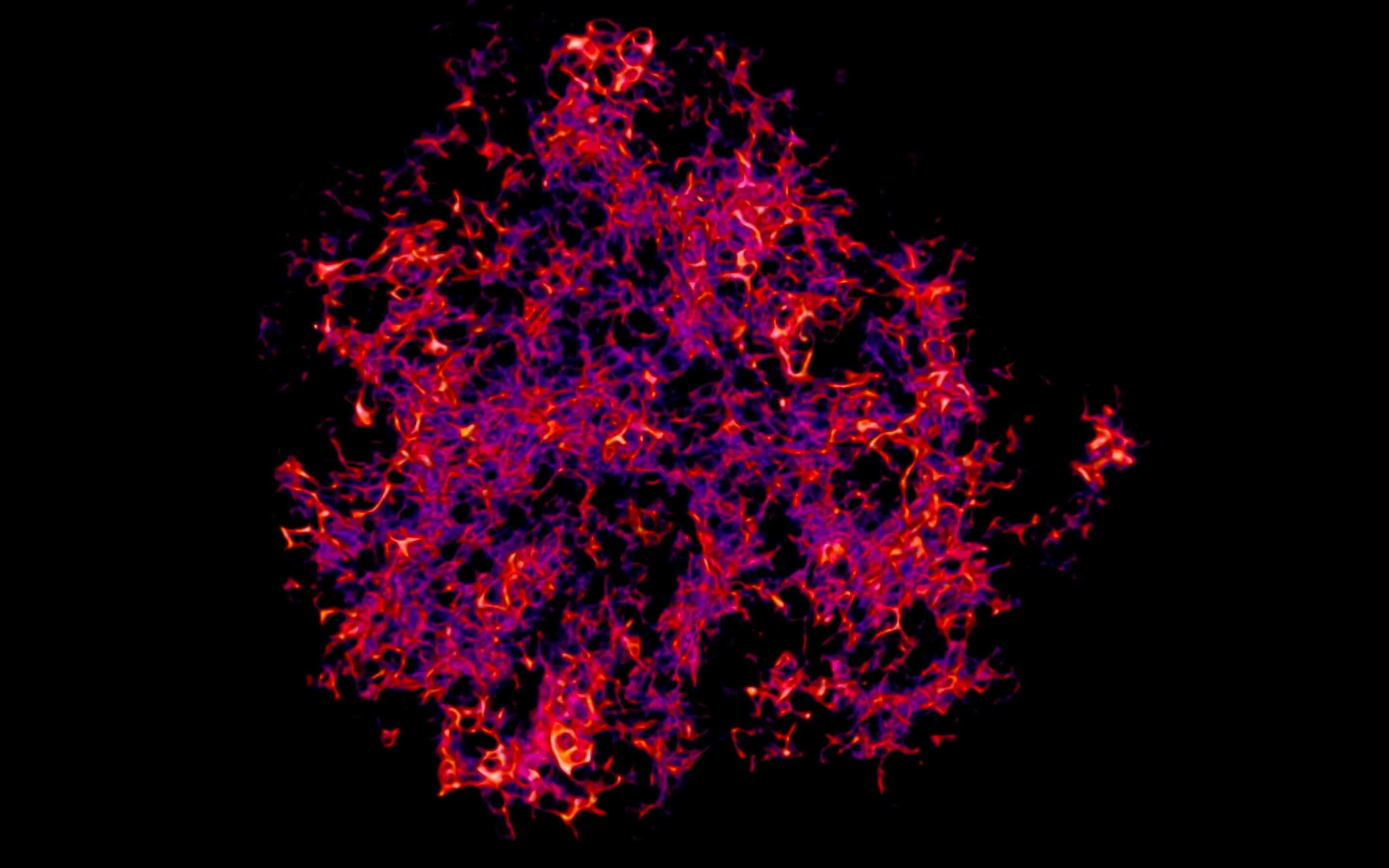
Intravital multiphoton microscopy showing T–B cell contacts in the germinal center (Video S3).
What do the human data tell us?
The study also examined human lymphoid tissue and tumors. Spatial transcriptomic data from human tonsils placed PD-L1 expression in regions with FDC signatures, consistent with the original observation in human FDCs. In tumor samples, analyses of renal-cell carcinoma tertiary lymphoid structures (TLS, organized immune-cell clusters that can form within tumors) also associated FDC signatures with CD274, the gene encoding PD-L1.
The team then looked for SOSTDC1-associated TFH cells in human cancer datasets. In triple-negative breast cancer, patients who responded to a treatment regimen containing the anti-PD-L1 antibody atezolizumab had lower SOSTDC1-positive TFH signatures than nonresponders. The signature did not distinguish responders from nonresponders in patients who received chemotherapy without anti-PD-L1 treatment. In a separate analysis of metastatic urothelial cancer patients treated with atezolizumab, a higher estimated abundance of SOSTDC1-positive TFH cells was associated with poorer overall survival, whereas a higher overall TFH-cell estimate was associated with better survival.
These findings are intriguing, but they are associations in patient datasets, not proof that SOSTDC1-positive TFH cells cause resistance to checkpoint blockade. They do suggest that the impact of PD-L1 blockade on tumors may depend partly on the state of the follicular T-cell and B-cell response, not only on conventional antitumor T cells.
What the study does (and does not) show
The central mechanistic evidence comes from mouse genetics and perturbation experiments. The human tumor analyses extend the relevance of the pathway, but they do not establish that the same causal mechanism drives clinical treatment response. There are also open questions: other cells in germinal centers can express PD-L1, the contribution of PD-L2 remains unresolved, and the precise molecular signals downstream of PD-1 that regulate TFH-cell Sostdc1 are not yet known. The study also does not distinguish whether Sostdc1 promotes TFR development from conventional regulatory T-cell precursors, from TFH cells, or from both in the tested setting.
Those limitations matter because they keep the interpretation appropriately focused. The paper does not show that PD-L1 is universally beneficial, or that blocking PD-L1 will always weaken antibody responses. It shows that PD-L1 expressed by germinal-center stromal cells (especially FDCs in the autoimmune models studied) can support humoral immunity through a pathway that differs from its familiar role in suppressing cytotoxic T-cell responses.
A long journey from observation to function
For me, this paper brings a long-running question full circle: from noticing PD-L1 and PD-L2 on human FDCs in 2018, to reporting the observation in 2021, to now testing what PD-L1 does in germinal centers. It took years, many experiments, and a truly Herculean effort by Elliot Akama-Garren to get here.
The work gives us a new way to think about the germinal-center “checkpoint”. FDC PD-L1 can help maintain the balance between helper and regulatory follicular T cells, support stable B-cell–TFH interactions, and promote antibody responses. In autoimmune mouse models, the same activity can also help autoreactive germinal centers expand and admit new B-cell clones. In cancer, the human data raise the possibility that SOSTDC1-positive TFH cells may be relevant to responses to PD-L1 blockade, a hypothesis that will need direct testing.
Read the paper: PD-L1 expression in germinal centers promotes humoral immunity by restricting T follicular regulatory cell differentiation.
Elliot, congratulations and thank you for making this project a success!